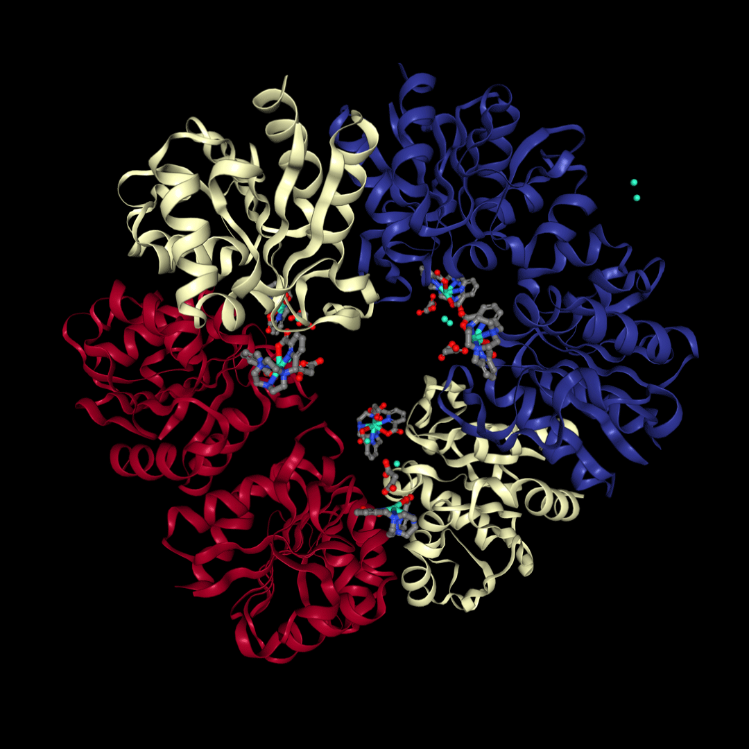
If DNA and RNA are the blueprints of life, proteins are the molecular machines working in its factory.
Proteins provide a multitude of functions within the living cell and in tissues. By schematizing, a protein is like a pearl necklace, a chain of small molecules called amino acids. Predicting how the necklace of several hundred “pearls” will fold into space is now a real technological challenge. But it is crucial because there is a direct link between the 3D structure of the protein and its biological function.
The use of proteins as therapeutics or targets for therapeutics to treat disease, continues to drive protein research. Key application areas include protein-protein interaction studies, drug screening, target identification, biomarker discovery, protein therapeutics, diagnostic tests, and disease monitoring. Determining the structure of proteins, is thus a crucial step for the understanding of biological processes.
Thanks to synchrotron radiation, biological crystallography (bio-crystallography) aims to produce precise three-dimensional “architectural” models of biological molecules. With such models in hand, it facilitates the understanding of biological processes – for example, the way proteins and other molecules behave in cells – or to design drugs that will target their functions. The two main limitations of the method are (i) obtaining suitable crystals, and (ii) phasing the diffraction data

